Specialists who treat this condition
Mr Mark Batterbury
Mr Nicholas Beare
Mr Austin McCormick
Prof Ian Pearce
There are many different minor lesions that may affect the eyelid. Most are a cyst or papilloma.
A cyst is caused by a gland, which has become blocked but continues to produce fluid or oil. This collects under the skin as a spherical lump.
A papilloma is a warty fleshy growth of skin, also known as a skin tag. They are benign and not a form of skin cancer.
Cysts and papillomas may cause irritation, watering of the eye and some people do not like the appearance.
Many people ask for them to be removed surgically. Some people are worried that the lump may be cancerous and removing it enables a pathologist to examine it microscopically and exclude skin cancer.
It is important not to ignore lesions if they are changing as this may be a sign that the are cancerous.
Chalazion
A chalazion is a small pea sized lump in the eyelid. It is due to blockage and infection in a meibomian gland in the eyelid.
The meibomian glands sit within the cartilage of the eyelid and open up in a row behind the eyelashes. They produce oil for the tear film.
At first the margin of the eyelid may become red. This progresses to form a lump with redness and swelling of the eyelid skin. This acute phase usually passes within a couple of weeks leaving a small, non-reddened lump called a chalazion as seen above.
Many meibomian gland infections do not develop into a chalazion and will simply settle spontaneously.
As soon a red mark on the eyelid is seen intensive hot compresses should be carried out to try to prevent the formation of a chalazion. 6 times a day for 5 minutes at a time.
Even once a chalazion has formed most will settle within 3 months without treatment.
If it does not settle or if it is particularly large an incision and curettage may be carried out.
This is a minor surgical procedure carried out in theatre or treatment room.
Local anaesthetic is injected into the eyelid. An incision is made on the inside of the eyelid allowing pus to be drained. No sutures are required.
An eye pad may be placed to be kept on until you return home.
You will be given antibiotic ointment to be put in the eye for 5 days post operatively.
Cyst of Moll
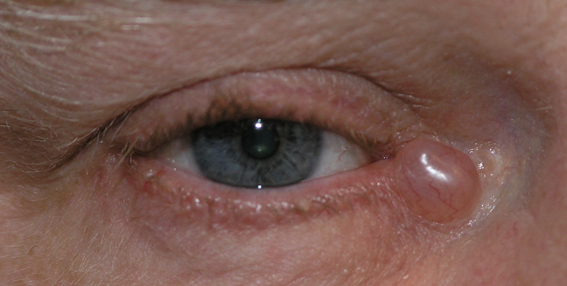
This is a benign cyst of a gland of Moll. This is a small gland that sits at the base of an eyelash.
It is a clear fluid filled cyst.
It can be treated with surgical excision. This may be carried out in a treatment room under local anaesthetic.
Your consultant will remove the cyst in its entirety to reduce the risk of recurrence.
After surgery ointment will be applied 3 times a day for two weeks. The area where the cyst was will form a small scab and then heal over the next two weeks.
Cyst of Zeiss
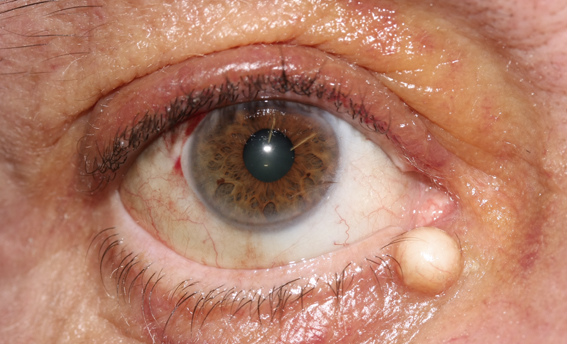
Acyst of zeiss is a benign cyst arising from a gland of Zeiss.
Glands of Zeiss are found at the base of eyelashes along side glands of Moll.
Instead of clear fluid they are filled with yellow oily secretions.
They may be excised under local anaesthetic in a treatment room.
Afterwards you will need to apply antibiotic ointment 3 times a day for two weeks during which time the small scab left where the cyst used to be will heal.
Sebaceous cyst
This is a benign skin cyst
A sebaceous cyst occurs in sebaceous glands in the skin. They can occur anywhere on the body.
Sebaceous glands produce oil for the skin hence the yellow colour of the cyst
They can be excised under local anaesthetic in a treatment room.
After surgery small dissolvable sutures may be used or there may be a small scab that will heal over within 2 weeks.
Antibiotic ointment should be applied 3 times a day for 2 weeks.
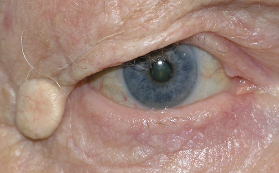
Papilloma
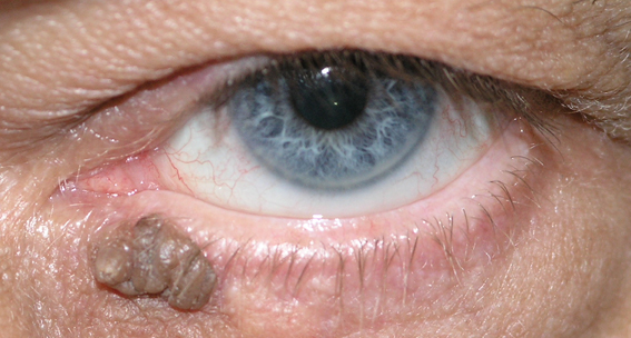
Papillomas are benign overgrowths of skin. They are also known as skin tags.
They may be pink or pigmented as above.
They are caused by viral infection with human papilloma virus. Many people are infected with this virus without knowing it. It causes warty growths on the skin.
They may be excised surgically under local anaesthetic in a treatment room.
After surgery a small scab may form. Antibiotic ointment should be applied to the area 3 times a day for 2 weeks
When is a lesion benign and when is it malignant?
It is important not to mistake a benign lesion for a skin cancer. If there is any doubt, the excised lesion will be sent to a pathologist who will examine it under a microscope and determine the diagnosis.
Your consultant will ask you how the lesion arose and he will examine you carefully. It is usually possible from this history and examination to determine if the lesion is benign or malignant.
A malignant lesion grows more rapidly than a benign one. It may ulcerate or bleed. It may from a crust in the centre. It may be painful. The normal shape of the eyelid may change and eyelashes may be lost. Any of these features are suspicious and a biopsy to determine the diagnosis should be carried out first.
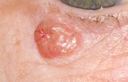
LEFT: All three of these lesions are malignant. This first one is a basal cell carcinoma. It has shiny rolled edges with a pit or crater in the centre. There are dilated blood vessels at the edges.
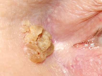
MIDDLE: This squamous cell carcinoma has a hard crust on the surface. This sometimes comes off revealing an ulcer underneath. The crust reforms and the ulcer grows.
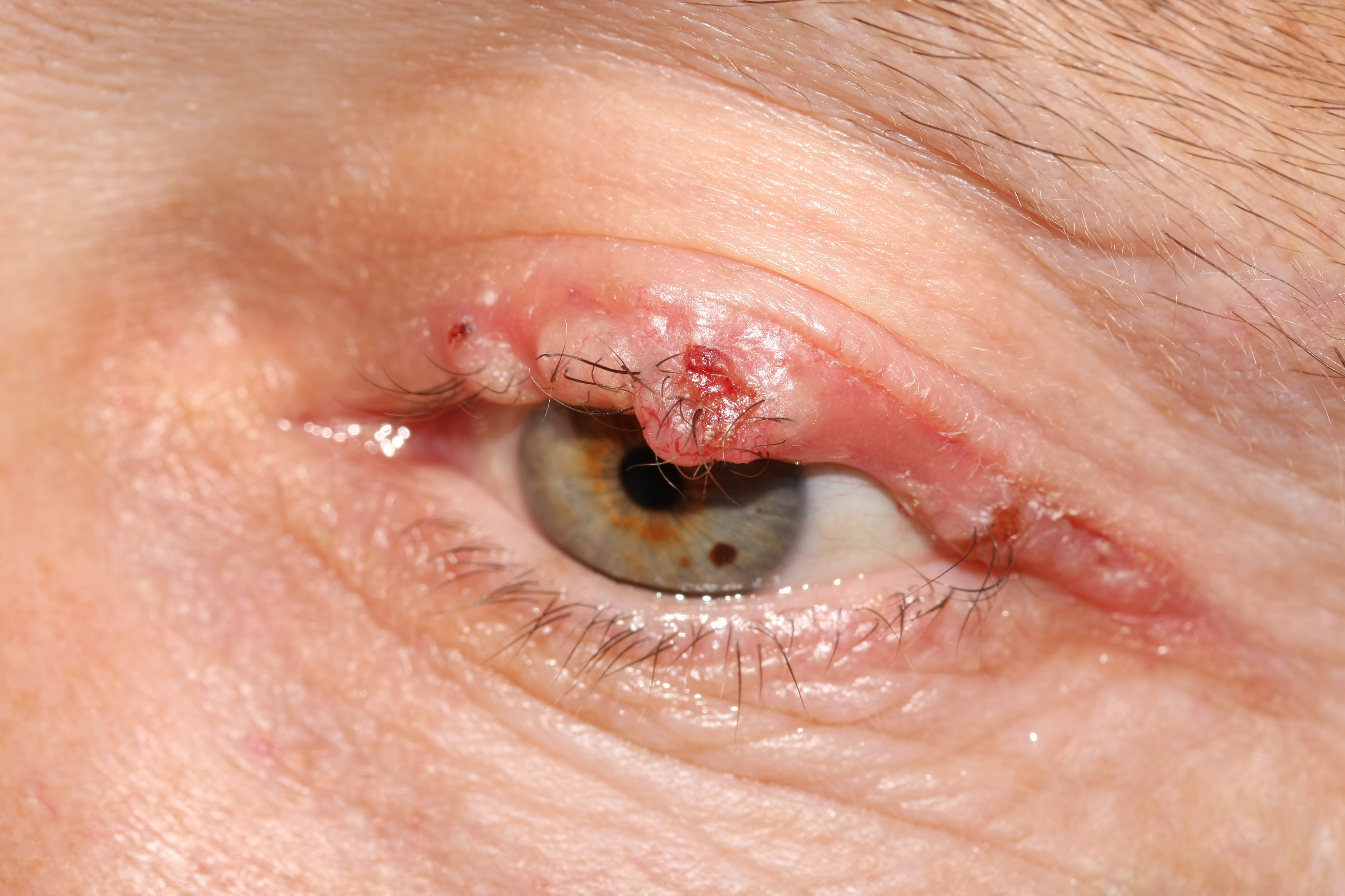
RIGHT: This upper lid basal cell carcinoma has spread along the margin of the eyelid. The normal anatomy has been lost: eyelashes have gone and the normal square edge to the lid is replaced with a rounded and notched one.